Small device contains wells to let small bits of tissue grow, develop, and be studied in real time
[ Source: American Institute of Physics | April 06, 2021 ]
Scientists from MIT and the Indian Institute of Technology Madras have grown small amounts of self-organizing brain tissue, known as organoids, in a tiny 3D-printed system that allows observation while they grow and develop. The work is reported in Biomicrofluidics, by AIP Publishing.
Current technology for real-time observation of growing organoids involves the use of commercial culture dishes with many wells in a glass-bottomed plate placed under a microscope. The plates are costly and only compatible with specific microscopes. They do not allow for the flow or replenishment of a nutrient medium to the growing tissue.
Recent advances have used a technique known as microfluidics, where a nutrient medium is delivered through small tubes connected to a tiny platform or chip. These microfluidic devices are, however, expensive and challenging to manufacture.
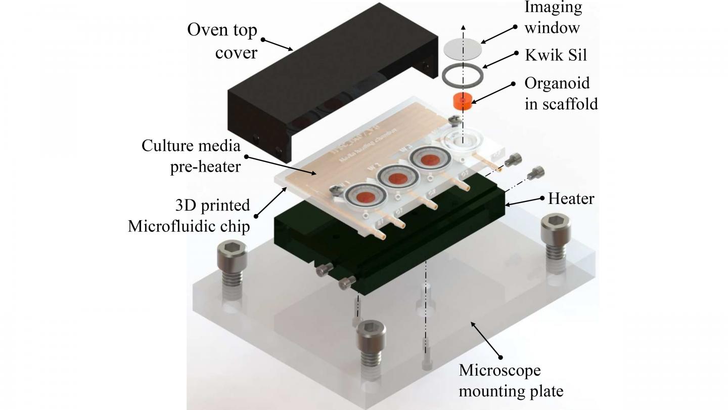
The current advance uses 3D printing to create a reusable and easily adjustable platform that costs only about $5 per unit to fabricate. The design includes imaging wells for the growing organoids and microfluidic channels to provide a nutrient medium and preheating that supports tissue growth.
A biocompatible type of resin used in dental surgery was used for the 3D-printed device. The printed chip was cured by exposing it to UV light, then sterilized before live cells were placed in the wells. After sealing the top of the wells with a glass slide, the nutrient medium and drugs for use in the study were added through small inlet ports.
“Our design costs are significantly lower than traditional petri dish- or spin-bioreactor-based organoid culture products,” said author Ikram Khan. “In addition, the chip can be washed with distilled water, dried, and autoclaved and is, therefore, reusable.”