Engineers and neuroscientists working to advance multiphoton microscopy are pushing the depths, speeds, and scales of in vivo imaging.
[ Source: Neff, E.P. Set lasers to image. Lab Anim 49, 245–248 (2020). https://doi.org/10.1038/s41684-020-0623-0 | August 13th, 2020 ]
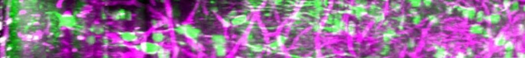
“Murat Yildirim, a postdoctoral fellow working with Peter So and Mriganka Sur at MIT who recently built a 3P microscope that could record evoked neural activity in all 6 layers of the mouse visual cortex and into the white matter beneath, is looking to apply the technology not just in mice, but also in smaller animals in the future: flies, worms, even Hydra, as the genetic toolboxes for these animals expand. A 3P microscope may still only reach a short distance into the cortex of a large macaque, but it could capture larger volumes of the nervous system in smaller animals. In zebrafish for example, a 3P system made it through the opaque skulls of adult fish to image the entire forebrain and deep into the cerebellum, reaching depths of 750 µm.
But with great power comes great responsibility: to compensate for the lack of efficiency with added photons, researchers need to push greater power into a tighter time space—from microseconds to femtoseconds, says Podgorski; that runs the risk of photodamaging tissue. “As neuroscientists and as other scientists are pushing the boundaries on how much they want to measure in a given subject, we’re using higher and higher laser powers, we’re putting more energy into the brain, and at some point you start to do damage and you need to know when that’s occurring,” he says.
Those thresholds can vary between organisms: while a mouse brain might handle 250 milliwatts of power, a more sensitive fly might only tolerate 15 to 20 milliwatts. ”You have to know in advance how deep you want to image, and then you pick the technology to suit the experiment,” he says.
As researchers continue to push the limits of 2P & 3P microscopes—their depth, speed, and scale, plus the development of new and better indicators (Box 1), some questions remain about accessibility. “It’s very hard to share complicated instruments,” says Podgorski. “It’s so satisfying when we make a new fluorescent indicator and someone wants to use it and we just put it in the mail. We can’t do that with microscopes.”
There are moves in two directions in the field: to make instruments cheaper, and to set up observatories. The Advanced Imaging Center at Janelia funds researchers to in and use its instruments, for example. Lasers, particularly 3P versions, aren’t cheap; the commercial microscopes to go with them can sometimes lack flexibility, while specialized scopes can be tricky for those without engineering backgrounds to construct—Yildirim likens it to a restaurant chef, who must consistently serve a wide range of tastes vs. the family cook who can tweak and experiment at home.
“In engineering you can develop many, many microscopes,” he says. But an important question remains: how can you transfer those to neuroscience labs? Engineers need to demonstrate that their microscopes are more than just proof of the latest concept, but that they can stand up to repeated, long-term use that scientists need to perform their experiments, he says.”