
[Source: David Orenstein | The Picower Institute for Learning and Memory | August 07, 2025]
By combining several cutting-edge imaging technologies, a new microscope system could enable unprecedentedly deep and precise visualization of metabolic and neuronal activity, potentially even in humans
Both for research and medical purposes, researchers have spent decades pushing the limits of microscopy to produce ever deeper and sharper images of brain activity, not only in the cortex but also in regions underneath such as the hippocampus. In a new study, a team of MIT scientists and engineers demonstrates a new microscope system capable of peering exceptionally deep into brain tissues to detect the molecular activity of individual cells by using sound.
“The major advance here is to enable us to image deeper at single-cell resolution,” said neuroscientist Mriganka Sur, a corresponding author along with mechanical engineering Professor Peter So and principal research scientist Brian Anthony. Sur is the Paul and Lilah Newton Professor in The Picower Institute for Learning and Memory and the Department of Brain and Cognitive Sciences at MIT.
In the journal Light: Science and Applications, the team demonstrates that they could detect NAD(P)H, a molecule tightly associated with cell metabolism in general and electrical activity in neurons in particular, all the way through samples such as a 1.1 mm “cerebral organoid,” a 3D-mini brain-like tissue generated from human stem cells, and a 0.7 mm thick slice of mouse brain tissue.
In fact, said co-lead author and mechanical engineering postdoc W. David Lee, who conceived the microscope’s innovative design, the system could have peered far deeper but the test samples weren’t big enough to demonstrate that.
“That’s when we hit the glass on the other side,” he said. “I think we’re pretty confident about going deeper.”
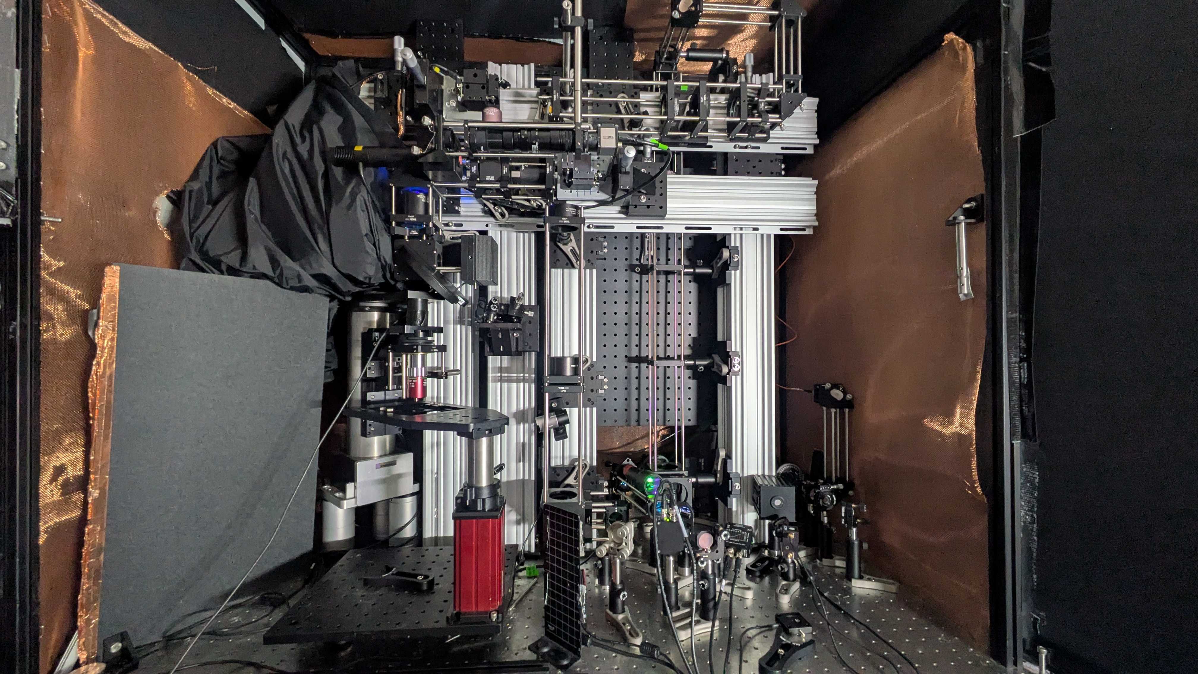
Still, a depth of 1.1 mm is more than five times deeper than other microscope technologies can resolve NAD(P)H within dense brain tissue. The new system achieved the depth and sharpness by combining several advanced technologies to precisely and efficiently excite the molecule and then to detect the resulting energy all without having to add any external labels, either via added chemicals or genetically engineered fluorescence.
Rather than focusing the required NAD(P)H excitation energy on a neuron with near ultraviolet light at its normal peak absorption, the scope accomplishes the excitation by focusing an intense, extremely short burst of light (a quadrillionth of a second long) at three times the normal absorption wavelength. Such “three-photon” excitation penetrates deep into tissue with less scattering by brain tissue because of the longer wavelength of the light (“like fog lamps,” Sur said). Meanwhile, though the excitation produces a weak fluorescent signal of light from NAD(P)H, most of the absorbed energy produces a localized (~10 microns) thermal expansion within the cell, which produces sound waves that travel relatively easily through tissue compared to the fluorescence emission. A sensitive ultrasound microphone in the microscope detects those waves and, with enough sound data, software turns them into high-resolution images (much like a sonogram does). Imaging created in this way is “three-photon photoacoustic imaging.”
“We merged all these techniques—three-photon, label-free, photoacoustic detection,” said co-lead author Tatsuya Osaki, a research scientist in The Picower Institute in Sur’s lab. “We integrated all these cutting-edge techniques into one process to establish this ‘Multiphoton-In and Acoustic-Out’ platform.”